Neuroscience study published by researchers of Oregon Health & Science University, Portland, Oregon, USA suggests widespread dysconnectivity in ADHD brain.
Objectives
Researchers aimed to address the challenges of identifying replicable neuroimaging correlates of ADHD by focusing on the cumulative effects of resting-state connectivity across all brain networks. In simple terms, this study looked at how brain connectivity, or how different parts of the brain communicate with each other, is related to symptoms of attention-deficit/hyperactivity disorder (ADHD).

The researchers used brain scans from a large number of people to create a score that represents the overall level of connectivity in the brain and how it relates to ADHD symptoms: a large, multi-site sample from the Adolescent Brain Cognitive Development (ABCD) study and an independent Oregon-ADHD-1000 case-control cohort to construct and validate a polyneuro score (PNRS) representing cumulative, brain-wide, ADHD-associated resting-state functional connectivity.
Results
The results showed a significant association between the ADHD PNRS and ADHD symptoms in both cohorts after accounting for relevant covariates. This suggests that differences in brain connectivity may play a role in the development of ADHD.
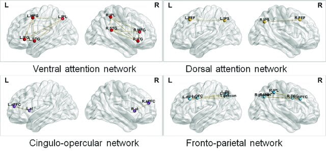
The most predictive PNRS involved all brain networks, with the strongest effects concentrated among connections involving the default mode and cingulo-opercular networks.

Furthermore, in the longitudinal Oregon-ADHD-1000 cohort, non-ADHD youth had significantly lower PNRS than children meeting ADHD diagnostic criteria at multiple time points.
However, the PNRS did not mediate polygenic risk for ADHD.
The study highlights the widespread dysconnectivity in ADHD and underscores the promise of the PNRS approach for improving reproducibility in neuroimaging studies and understanding the complex relationships between brain connectivity and behavioral disorders.
The research was supported by the National Institutes of Health and emphasized the significance of using large samples for improving reproducibility in neuroimaging studies.
The findings contribute to a better understanding of the cumulative effects of resting-state connectivity across brain networks in relation to ADHD symptoms, thereby advancing the understanding of the neurobiological underpinnings of the disorder.
References
Freedman, Lidan, et al. “Greater Functional Connectivity Within the Cingulo-opercular and Ventral Attention Networks Is Related to Better Fluent Reading: A Resting-state Functional Connectivity Study.” NeuroImage: Clinical, vol. 26, Jan. 2020, p. 102214, doi:10.1016/j.nicl.2020.102214.
Mooney, Michael A. “Cumulative Effects of Resting-state Connectivity Across All Brain Networks Significantly Correlate With ADHD Symptoms.” Journal of Neuroscience, Jan. 2024, doi:10.1523/JNEUROSCI.1202-23.2023.

Leave a Reply